
An ongoing dialogue on HIV/AIDS, infectious diseases,
August 30th, 2015
(Not) Doing the Retinal Exam, and the Importance of Acknowledging Limitations
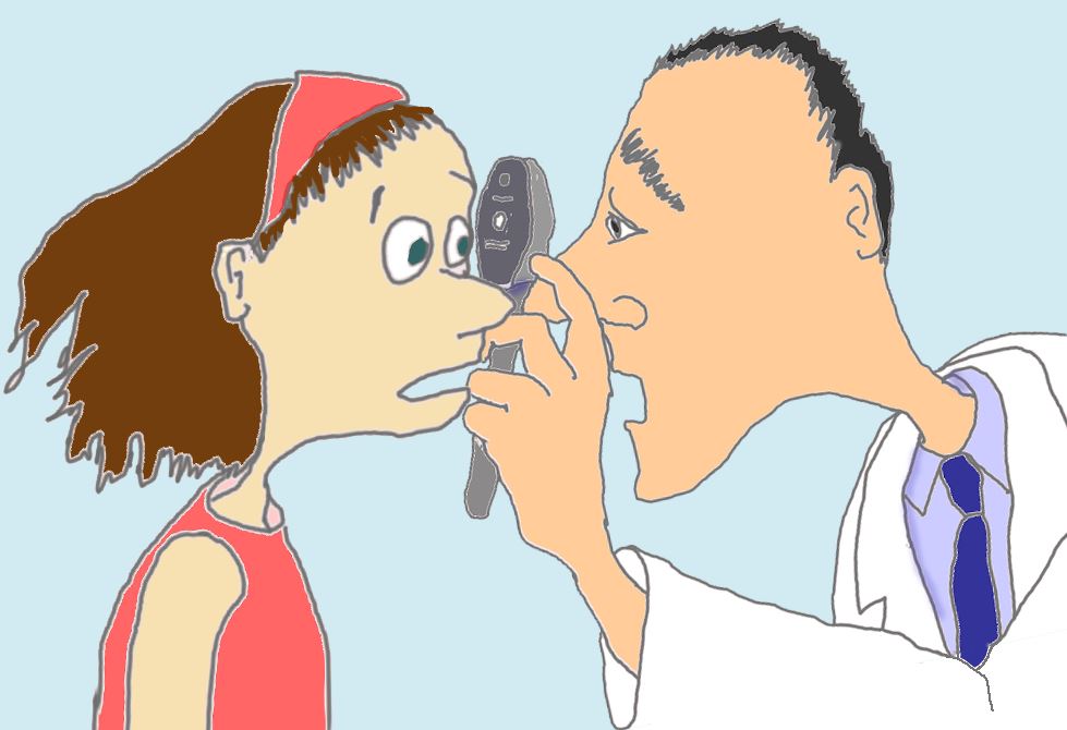 This past week, the New England Journal of Medicine released one of its excellent instructional videos, detailing how to do direct ophthalmoscopy to examine the retina.
That’s the use of one of those hand-held gizmos — an ophthalmoscope, see picture on the right — to look at the back of the eye.
As usual, the video was clear, succinct and professionally done. A great resource for clinicians, young and old.
But here’s a confession — the video could be a nominee for this year’s Oscar for Best Picture, it wouldn’t help me a bit. Because I’m absolutely horrible at this procedure, always have been.
I’m as likely to see something important in someone’s eyes using an ophthalmoscope as I am using a shoe, an avocado, or a tennis racket (to choose three random things that happen to be in the room right now as I write this).
The inability to do a particular part of the physical examination creates some uncomfortable moments, in particular during medical school when learning the skill. Asked to acknowledge that yes, we do in fact see/hear/feel what we’re being taught to see/hear/feel, medical students face a dilemma when failing to do so. In essence, there are three options:
- Tell the truth: “I can’t see the venous pulsations” — and ask the instructor for more guidance right there on the spot.
- Lie: “Oh yeah, the optic disc margin is sharp” — this gives the teacher a sense that you’ve mastered the technique, but it’s basically cheating and a very bad idea in clinical medicine, for innumerable obvious reasons.
- Silence: Be quiet on the matter, blending into the background and allowing the class to move on — and remain hopeful that you’ll pick up the skill later with more practice.
I wish I could say I had the maturity to go with #1, but in fact #3 was the route I chose.
Even if I had chosen #1, however, I’m not sure it would have made much of a difference. The reason I can’t do this procedure is because my own eyes are so bad — which, for a variety of reasons having to do with the optics of it, make direct ophthalmoscopy virtually impossible.
Trust me on this one — it’s not just an excuse. (Of course it is.) The right eye is particularly useless, which means I’d have to use my left eye to look into a patient’s right eye. If you imagine doing this, it sets up a very uncomfortable nose-to-nose encounter with your patient — highly unhygienic and socially weird, a reason right there not even to try it.
In the face of limitations we have as doctors, we invariably come up with rationalizations to make ourselves feel better. And here are mine about this particular deficit:
- Visual complaints are serious business — wouldn’t this deserve a referral to a real pro, an ophthalmologist? You can be sure that in the CMV retinitis days, my threshold for referral was very very low.
- Similarly, let’s say I found something incidentally on ophthalmoscopy — certainly this would warrant having an ophthalmologist see the patient to confirm, right? What non-eye doctor would be 100% sure saying that what they visualized with their crude scope is benign?
- Direct ophthalmoscopy isn’t even the best technique to look at the retina — the indirect method is far superior, preferred by all retina specialists.
- There are many other things in medicine I can’t do (replace heart valves, biopsy colons, do immunohistochemical stains, interpret EEGs, remove thyroid tumors) — this is just one more.
- I may not be able to see someone’s retina (especially their right retina, see above), but I’m pretty good at listening to heart murmurs, if I do say so myself. Hey, I once noticed that a patient with endocarditis developed aortic insufficiency before the cardiologist did. Granted, she hadn’t seen him that day yet, but still …
In all seriousness, the real reason for this confession is that I’m pretty sure we all have limitations. Why else would the orthopedists consult us ID doctors for essentially every infectious complication on their patients, no matter how simple? After all, before their residency, these pre-orthopods were some of the smartest medical students — did they suddenly lose their brains when they began doing knee replacements?
Not a chance. It’s simply that at some point, it makes much more sense to acknowledge these limitations — and move on — than to pretend they don’t exist, or even worse, to fake it.
9 Responses to “(Not) Doing the Retinal Exam, and the Importance of Acknowledging Limitations”

Paul E. Sax, MD
Contributing Editor
NEJM Journal Watch
Infectious Diseases
Biography | Disclosures | Summaries
Learn more about HIV and ID Observations.
 NEJM Journal Watch — Recent Infectious Disease Articles
- Innovative Pairing of Beta-Lactamase Inhibitors to Combat Multidrug-Resistant Gram-Negative Pathogens
- Ceftobiprole Joins the Beta-Lactam Squad Against MRSA Infections
- Sulbactam-Durlobactam for Treating Patients with Acinetobacter baumannii Infections: Do MICs Matter?
- Antibiotic Options Against Enterobacterales That Produce Wild-Type AmpC β-Lactamases
- Quorum Sensing in Pseudomonas aeruginosa — An Overlooked Treatment Target?
May I recommend “University Days” by James Thurber as a few paragraphs of similar ocular difficulties, i.e. trouble seeing little bitty things magnified through small glass orifices (orifi?)
Enjoy, I hope.
Sue V
Hi Sue,
I am a huge Thurber fan — My Life and Hard Times is probably one of my favorite books of all time!
Paul
Hi Dr. Sax,
Thank you for the great post! I remember in medical school having to repeatedly tell my attending that I did not hear the crackles and it took weeks for me to get it. It was embarrassing but I had a great attending who valued teaching and never made me feel bad. Unfortunately, there is a lot of pressure to “fake it,” even early on in medical training. Also, there is a lot of pressure to fake knowledge. Numerous times in rounds I recall asking the person next to me a question and they didn’t know the answer, but everyone was too afraid to speak up so just went with option #3.
Appreciate your post and bringing this to people’s attention.
Jodie
Dear Dr. Sax,
I saw venous pulsations for the first time as a junior medical student. Heidi, our miniature schnauzer, was paralyzed when I looked at her eyes with my ophthalmoscope. As if she had been frozen in carbonite. (Extra points will be awarded if you catch that allusion.) I could look at her retina for several minutes and she would not move a muscle. Finally saw venous pulsations there. Thanks for reminding me of a pleasant memory.
Sincerely yours,
Bryan Burke MD
Well said. I’m also very bad at looking at the retina. I wonder how Dr. Abraham Verghese would react to this post, given his efforts with the “Stanford 25.”
http://stanfordmedicine25.stanford.edu/the25/
This is #12
Thanks, Phil!
One day I’ll have the guts to write what I really think about the “cult” of the physical examination, but not quite yet.
Paul
Oddly enough, my doctor does in fact use an avocado to examine my eyes. It works surprisingly well.
Old; old school internist “until I saw the light”; ophthalmologist and glaucoma specialist, I well know the sad limitations of medical schools’ teaching of eye diseases and the eye exam. But I wouldn’t “give in” to the excuse that difficulty with a fundus exam prevents a decent eye examination — crucial to the H&P.
Here are my observations as to what things are important:
1. Way too much emphasis by attendings on A/V nicking/ratio, H&E, etc.
2. Seeing and understanding the convex iris (vs. flat vs. concave) viewed side on to identify the potential for angle closure caused by nature, uncommonly by dilating gtt.(v.i.). One can argue that a precipitated angle closure in a medical setting is better than closure spontaneously. Prompt laser iridotomy is practically curative.
3. Using the well-maintained direct ophthalmoscope to your best advantage, even if you don’t understand + vs. -, to examine the anterior segment as well as the fundus ( you may then understand why you can’t see the fundus, because of patient’s optical media, usually cataract, and then be assured that cataract is a legitimate diagnosis.
4. Ask your friendly ophthalmology resident about an afferent pupillary defect, what it means, and how to examine pupils properly. He also has access to “practice eyes” to learn fundoscopy.
5. Understand the prevalence of glaucoma — if you are not detecting glaucoma in a significant # of adult patients, say 1-2/100 on optic nerve exam alone, you are missing potential blindness.
6. Finally, check vision and instill dilating drops early in the encounter (usual caveats – head injury, marked farsightedness, read thick convex glasses or contact lenses, etc.) Fundus exam comes last. Remove your glasses to examine the patient (or not, depending on which way you can see better, but the closer “pupil-to-pupil” the exam, and the larger the pupil, the better the view). (Personal to Dr. Sax — sorry about your vision, but you can use your OS to patient OD if he is recumbent.)
7. Yes, refer early, to the ophthalmologist (the MD).
8. BTW, “PERRLA” is a prima facie joke. I saw convergence tested but rarely the pupils’ reflex associated with convergence and accommodation.) A good pupil exam takes seconds. An afferent pupillary defect is very common, very commonly missed, and often crucial to a diagnosis.
DJJ
As a hospitalist at a psychiatric hospital these days, and being old and honest, I simple put, ‘fundi not visualized’. Also, as DJJ stated above, that PERRLA thing…. I can’t believe how commonly I see fairly small pupils that don’t constrict with light, especially in patients over mid 50s. So, my honest eye exam is often, “pupils equal, about 2-3 mm, NR to light”. Period. In 41 years of medicine, I think the only time I’ve seen pupils that weren’t round is after surgery or injury, ie, very uncommon – to the point of not bothering to mention it. And who reads this stuff anyway? Now, anisocoria is far more common and interesting, but another finding about which there’s usually nothing to do. Except in undiagnosed Lyme patients….
jon