
July 25th, 2013
The Changing Landscape of Pulmonary Hypertension
John Ryan, MD
In the New England Journal of Medicine this week, Ghofrani et al. present two multicenter clinical trials on the role of a new oral pulmonary vasodilator in pulmonary hypertension. Riociguat is a soluble guanylate cyclase stimulator that promotes vasodilation in the pulmonary circulation. Riociguat represents a new class of medicines available for pulmonary hypertension and therefore will change the landscape of therapeutic options available in this disease process. However, there are a couple of aspects worth noting about these studies.
In the PATENT-1 trial, the researchers study the effects of riociguat in Group 1 PH, pulmonary arterial hypertension (PAH). Current treatment options available for this group are prostacyclins, endothelin receptor antagonists (ERA), phosphodiesterase 5 inhibitors (PDE5i), and calcium-channel blockers (the latter used only in those deemed reversible on right heart catheterization). Epoprostenol (prostacyclin) has been shown to improve survival in PAH but is administered via continuous intravenous infusion, and is thus limited in terms of ease of use as well as its cost. ERA and PDE5i have been shown to improve 6-minute walk distance, which is the same primary outcome used in PATENT-1. Most of these agents typically improve 6-minute walk distance by about 30 to 50 meters. In the PATENT-1 study, only 21% of treated patients had a functional improvement at 12 weeks. Therefore, it appears that riociguat is going to be another PAH drug of modest efficacy. Because of the near-equivalent outcomes, decision making regarding which agents to use as first-line or in combination therapy is subject to remarkable physician variability, depending on cost, familiarity, experience, access, and pharmaceutical company influence. To address this variability in care, primary outcomes that include endpoints such as mortality and hospitalization are becoming more standard in PAH trials, with decreasing emphasis being placed on 6-minute walk distance. Also, the trials in PAH remain small because it is still a rare disease with a prevalence of 6.6 cases/million adults. In addition, this study does not reflect the largest population of pulmonary hypertension in clinical practice, namely Group 2 PH (PH secondary to left heart disease), for which are still no dedicated therapies.
The CHEST-1 trial of riociguat in chronic thromboembolic pulmonary hypertension (CTEPH, Group 4 PH) is a unique study as it marks the first large, multicenter RCT of medical therapy to demonstrate marked clinical benefit in CTEPH. The primary outcome again is 6-minute walk distance, which improved by 46 meters in the treatment group compared with placebo. However, this surrogate clinical outcome is markedly inferior to the outcomes observed from pulmonary endarterectomy for treatment of this rare disease (accounting for about 2% of all cases of pulmonary hypertension). Therefore, it is important that patients identified as having CTEPH should still be presented with pulmonary endarterectomy as the first-line therapeutic option. In fact, to enter into CHEST-1, patients had to be deemed inoperable or had residual PH after undergoing pulmonary endarterectomy. The rare incidence of CTEPH coupled with the infrequent determination of inoperability should ultimately make riociguat a rarely prescribed agent for this group with PH.
July 25th, 2013
Vasopressin-Epinephrine Plus Corticosteroids Improve Neurologic Status After In-Hospital Cardiac Arrest – Implications for Out-of-Hospital Arrest?
Graham Nichol, MD MPH
Graham Nichol takes a close look at a recent randomized trial that examined whether combined vasopressin-epinephrine plus corticosteroids during CPR could improve survival and neurologic status in patients with in-hospital cardiac arrest. The trials results appear in JAMA.
Out-of-hospital and in-hospital cardiac arrest continue to be major public health problems in the U.S., with a combined burden of more than 500,000 fatalities annually.1,2 Many communities have not reported improved outcomes after out-of-hospital cardiac arrest (OHCA) for more than 30 years.3 Despite the apparent lack of progress, there is a more than fivefold variation in survival after treatment of OHCA across communities.4 This large variation suggests that cardiac arrest is a treatable condition that warrants further investigation.
Cardiac arrest involves sudden, global ischemia. Reperfusion occurs during cardiopulmonary resuscitation (CPR) and restoration of circulation, and is associated with a marked release of circulating inflammatory molecules including cytokines, activated complement and polymorphonuclear leukocytes, and endothelial cell adhesion molecules.5-8 After cardiac arrest, non-survivors demonstrate plasma interleukin-6 concentrations 20-fold greater than survivors,5 which is approximately 50-fold greater than normal human baseline values.9 In addition, patients with cardiac arrest have lower serum cortisol levels than those observed in other stress states.10 There is an association between serum cortisol and short-term outcomes in this population. Impaired response to corticotropin stimulation is not modified by application of induced hypothermia.11 These inflammatory and coagulation changes contribute to poor capillary perfusion, tissue ischemia, multi-organ dysfunction, and death.12-15 But many patients with early hemodynamic dysfunction that is reduced or treated do survive to have good neurologic outcomes.5,16 Therefore, treatments that decrease early mortality related to inflammation and intractable shock could increase the number of survivors with good outcomes.
Differences in how manual chest compressions are applied to patients with OHCA by EMS providers are associated with differences in outcomes. Greater chest compression fraction,17 greater depth of chest compression,18 greater chest compression rate,19 and briefer perishock pauses are associated with better outcomes.20 Thus, potential improvements in advanced cardiac life support should not be implemented at the expense of good basic care.
Previous studies provide conflicting estimates of the effect of intravenous epinephrine and vasopressors in patients with cardiac arrest. A randomized trial reported that use of intravenous drugs – including, but not limited to, epinephrine – improved survival to admission but not discharge compared with no intravenous drugs in patients with OHCA.21 Large effectiveness trials reported no survival benefit with vasopressin compared with epinephrine in patients with cardiac arrest.22-24 A recent large observational study derived from a Japanese OHCA registry suggested that use of epinephrine was not associated with improved outcomes compared with no epinephrine.25 However, concurrent analyses of overlapping datasets from the same registry suggested that early use of epinephrine was associated with improved outcomes compared with late or no epinephrine.26,27 Importantly, the only trial of epinephrine versus placebo in patients with OHCA did not achieve its intended sample size because of lack of EMS provider equipoise. Nonetheless, this trial reported that epinephrine significantly improved restoration of circulation and admission to hospital and tended to improve survival to discharge.28 Opinions of experts differ as to whether the consistent effect estimate and inconsistent significance observed in this trial reflects lack of power or lack of effect. In the absence of a randomized trial with adequate power to detect a significant difference in a clinically important outcome such as survival to discharge, I believe that the current standard of care during attempted resuscitation is to provide epinephrine as early as possible without disrupting provision of high-quality CPR.
Previously, a single-center, randomized, double-blind, placebo-controlled trial in Greece evaluated whether early immune modulation with or without vasopressors could improve outcomes in patients with in-hospital cardiac arrest with a first recorded rhythm of ventricular fibrillation.29 Eligible patients allocated to the intervention group received intravenously vasopressin 20 IU during the first five CPR cycles and methylprednisonlone 40 mg during the first CPR cycle (n=48), followed by treatment of post-resuscitation shock with hydrocortisone 300 mg daily for 7 days (n=27). Patients allocated to the control group received intravenous saline placebo during each time period (n=52 in resuscitation phase; n=15 in post-resuscitation phase). Concurrent interventions in both groups included intravenous epinephrine 1 mg during each CPR cycle. Intervention group patients, compared with controls, had greater restoration of circulation (81% vs. 52%, p=0.003) and survival to discharge (19% vs. 4%, p=0.02). Among patients with post-resuscitation shock (apparently defined as inability to maintain mean arterial pressure greater than 70 mm Hg without vasopressors after volume loading), those who received corticosteroids had improved survival to discharge compared with controls (30% vs. 0%, p=0.02). The significant and important benefits of vasopressin and corticosteroids during attempted resuscitation and corticosteroids after resuscitation were considered sufficiently promising as to warrant replication in a larger trial.
Now, results from that larger trial have been published in JAMA. The multicenter, randomized, double-blind, placebo-controlled trial, also in Greece, used similar methods to the pilot study to evaluate similar interventions, with the notable exception that the current study enrolled patients with in-hospital cardiac arrest who required vasopressors as opposed to patients with a first-recorded rhythm of ventricular fibrillation. The intervention patients (n=130), who were allocated to vasopressin and corticosteroids during attempted resuscitation, had greater restoration of circulation than the control patients (n=138), who received saline alone (84% vs. 66%, p=0.005). The intervention group also had greater functional neurologic status at discharge (14% vs. 5%, p=0.02). Among patients with post-resuscitation shock (n=149), intervention patients who received corticosteroids had greater functional neurologic status at discharge than did controls (21% vs. 8%, p=0.02).
The study investigators should be congratulated on demonstrating significant and important benefits associated with the use of vasopressors and corticosteroids during attempted resuscitation as well as corticosteroids after resuscitation. The present trial used multiple methods to confirm its internal validity, including deploying chromatography to confirm drug stability, monitoring concurrent care, and achieving a high rate of follow-up. A notable strength of this trial was the use of masked allocation, as unblinding can increase estimates of treatment benefit by about 50%.30 Use or non-use of blinding may explain discordant estimates of the effect of interventions in patients with OHCA.31,32
The study has a few limitations that may affect whether these results apply to patients with OHCA or in-hospital arrest in other settings. First, patients who were randomized but had circulation restored before study drug administration were excluded from the analysis, so the aggregate benefit of the intervention may be overestimated. Second, the intervention group received both vasopressin and corticosteroids during attempted resuscitation, so it is difficult to determine their relative contributions to improved outcome. Of note, patients in the control group frequently received open-label corticosteroids. But given the prior lack of incremental benefit of vasopressin in addition to epinephrine, it seems plausible that the primary benefit is attributable to immune modulation with corticosteroids. Third, patients with post-resuscitation shock were not re-randomized to corticosteroids versus placebo. Instead, patients who were allocated to vasopressin and corticosteroids during resuscitation and then subsequently developed shock received additional doses of corticosteroids. Thus, the relative importance of early versus later corticosteroids warrants further study.
There is large regional variation in the processes of care for patients with cardiac arrest.33,34 Importantly, interventions may have differential effects in patients with OHCA versus in-hospital cardiac arrest.35,36 Interventions that improve outcomes in one geographic region may not improve it in another due at least in part to differences in patient characteristics and processes of care.37,38 In addition, corticosteroids can impair myocardial healing in patients with acute myocardial infarction, which is commonly observed in patients with OHCA. The promising results of this trial warrant replication in a multicenter trial in centers that provide high-quality care to a high volume of patients with OHCA before adoption into clinical practice.
References
1. Go AS et al. Heart Disease and Stroke Statistics–2013 Update: A Report From the American Heart Association. Circulation. 2013 Jan 1;127(1):e6-e245.
2. Merchant RM et al. Incidence of treated cardiac arrest in hospitalized patients in the United States. Crit Care Med. 2011 Nov;39(11):2401-6.
3. Sasson C et al. Predictors of survival from out-of-hospital cardiac arrest: a systematic review and meta-analysis. Circ Cardiovasc Qual Outcomes. 2010 Jan;3(1):63-81.
4. Nichol G et al. Regional variation in out-of-hospital cardiac arrest incidence and outcome. JAMA. 2008 Sep 24;300(12):1423-31.
5. Adrie C et al. Successful cardiopulmonary resuscitation after cardiac arrest as a “sepsis-like” syndrome. Circulation. 2002 Jul 30;106(5):562-8.
6. Shyu KG et al. Concentrations of serum interleukin-8 after successful cardiopulmonary resuscitation in patients with cardiopulmonary arrest. Am Heart J. 1997 Sep;134(3):551-6.
7. Gando S et al. Alterations of soluble L- and P-selectins during cardiac arrest and CPR. Intensive Care Medicine. 1999;25(6):588-93.
8. Bottiger BW et al. Marked activation of complement and leukocytes and an increase in the concentrations of soluble endothelial adhesion molecules during cardiopulmonary resuscitation and early reperfusion after cardiac arrest in humans. Crit Care Med. 2002 Nov;30(11):2473-80.
9. Vgontas AN et al. I-6 and its circadian secretion in humans. Neuroimmunomodulation. 2005;12(3):131-40.
10. Schultz CH et al. A characterization of hypothalamic-pituitary-adrenal axis function during and after human cardiac arrest. Crit Care Med. 1993 Sep;21(9):1339-47.
11. Hekimian G et al. Cortisol levels and adrenal reserve after successful cardiac arrest resuscitation. Shock. 2004 Aug;22(2):116-9.
12. Lefer DJ and Granger DN. Oxidative stress and cardiac disease. Am J Med. 2000;109(4):315-23.
13. Anderson T and Vanden Hoek TL. Preconditioning and the oxidants of sudden death. Curr Opin Crit Care. 2003 Jun;9(3):194-8.
14. Chen Q et al. Production of reactive oxygen species by mitochondria: central role of complex III. J Biol Chem. 2003 Sep 19;278(38):36027-31.
15. Nolan JP et al. Therapeutic hypothermia after cardiac arrest: an advisory statement by the advanced life support task force of the International Liaison Committee on Resuscitation. Circulation. 2003 Jul 8;108(1):118-21.
16. Laurent I et al. Reversible myocardial dysfunction in survivors of out-of-hospital cardiac arrest. J Am Coll Cardiol. 2002 Dec 18;40(12):2110-6.
17. Christenson J et al. Chest compression fraction determines survival in patients with out-of-hospital ventricular fibrillation. Circulation. 2009 Sep 29;120(13):1241-7.
18. Stiell IG et al. What is the role of chest compression depth during out-of-hospital cardiac arrest resuscitation? Crit Care Med. 2012 Apr;40(4):1192-8.
19. Idris AH et al. Relationship Between Chest Compression Rates and Outcomes From Cardiac Arrest. Circulation. 2012 Jun 19;125(24):3004-12.
20. Cheskes S et al. Perishock pause: an independent predictor of survival from out-of-hospital shockable cardiac arrest. Circulation. 2011 Jul 5;124(1):58-66.
21. Olasveengen TM et al. Intravenous drug administration during out-of-hospital cardiac arrest: a randomized trial. JAMA. 2009 Nov 25;302(20):2222-9.
22. Stiell IG et al. Vasopressin versus epinephrine for inhospital cardiac arrest: a randomised controlled trial. Lancet. 2001 Jul 14;358(9276):105-9.
23. Gueugniaud PY et al. Vasopressin and epinephrine vs. epinephrine alone in cardiopulmonary resuscitation. N Engl J Med. 2008 Jul 3;359(1):21-30.
24. Wenzel V et al. A comparison of vasopressin and epinephrine for out-of-hospital cardiopulmonary resuscitation. N Engl J Med. 2004 Jan 8;350(2):105-13.
25. Hagihara A et al. Prehospital epinephrine use and survival among patients with out-of-hospital cardiac arrest. JAMA. 2012 Mar 21;307(11):1161-8.
26. Nakahara S et al. Association between timing of epinephrine administration and intact neurologic survival following out-of-hospital cardiac arrest in Japan: a population-based prospective observational study. Acad Emerg Med. 2012 Jul;19(7):782-92.
27. Hayashi Y et al. Impact of early intravenous epinephrine administration on outcomes following out-of-hospital cardiac arrest. Circ J. 2012;76(7):1639-45.
28. Jacobs IG et al. Effect of adrenaline on survival in out-of-hospital cardiac arrest: A randomised double-blind placebo-controlled trial. Resuscitation. 2011 Sep;82(9):1138-43.
29. Mentzelopoulos SD et al. Vasopressin, epinephrine, and corticosteroids for in-hospital cardiac arrest. Arch Intern Med. 2009 Jan 12;169(1):15-24.
30. Bero L et al. Factors associated with findings of published trials of drug-drug comparisons: why some statins appear more efficacious than others. PLoS Med. 2007 Jun;4(6):e184.
31. Aufderheide TP et al. Standard cardiopulmonary resuscitation versus active compression-decompression cardiopulmonary resuscitation with augmentation of negative intrathoracic pressure for out-of-hospital cardiac arrest: a randomised trial. Lancet. 2011 Jan 22;377(9762):301-11.
32. Aufderheide TP et al. A trial of an impedance threshold device in out-of-hospital cardiac arrest. N Engl J Med. 2011 Sep 1;365(9):798-806.
33. Zive D et al. Variation in out-of-hospital cardiac arrest resuscitation and transport practices in the Resuscitation Outcomes Consortium: ROC Epistry-Cardiac Arrest. Resuscitation. 2011 Mar;82(3):277-84.
34. Glover BM et al. Wide variability in drug use in out-of-hospital cardiac arrest: a report from the resuscitation outcomes consortium. Resuscitation. 2012 Nov;83(11):1324-30.
35. Bernard SA et al. Treatment of comatose survivors of out-of-hospital cardiac arrest with induced hypothermia. N Engl J Med. 2002 Feb 21;346(8):557-63.
36. Nichol G et al. Does induction of hypothermia improve outcomes after in-hospital cardiac arrest? Resuscitation. 2013 May;84(5):620-5.
37. Stiell IG et al. The Ontario trial of active compression-decompression cardiopulmonary resuscitation for in-hospital and prehospital cardiac arrest. JAMA. 1996 May 8;275(18):1417-23.
38. Plaisance P et al. A comparison of standard cardiopulmonary resuscitation and active compression-decompression resuscitation for out-of-hospital cardiac arrest. French Active Compression-Decompression Cardiopulmonary Resuscitation Study Group. N Engl J Med. 1999 Aug 19;341(8):569-75.
July 23rd, 2013
Funding for Landmark Framingham Heart Study Slashed by $4 Million
Larry Husten, PHD
One of the most important studies in the history of medicine will be sharply curtailed as a result of the federal budget cuts.
The landmark Framingham Heart Study (FHS) has been told by the National Heart, Lung and Blood Institute (NHLBI) that it will lose $4 million of its funding. The cut represents 40% of its NHLBI funding and is a direct consequence of the sequester, the automatic cuts in the federal budget.
The FHS employs about 90 people and receives some funding from other sources, including other divisions of the National Institutes of Health. According to a statement posted by the study on its website, the cuts will lead to the loss of 19 clinical and administrative jobs as well as “reductions in clinic exams and lab operations.” In addition, the FHS said:
Research will continue, but will be affected by the planned elimination of the examinations, which also are used in ancillary studies. The FHS will continue to remain in contact with its participants regarding future exam cycles.
We are working with the NHLBI leadership and with other potential funding sources to help sponsor the research, fill the funding gap and thus save these jobs and cover the costs of the previously planned clinical examinations and laboratory studies.
The FHS is administered by the Boston University Medical School. Future funding of the study is also threatened.
The FHS started in 1948 and has continued to study the original study participants and their descendants. The study — which coined the term “risk factor” — helped identify and understand the contribution to cardiovascular disease of cholesterol and other lipids, high blood pressure, smoking, obesity, diabetes, and many other factors.
July 22nd, 2013
Studies Raise Questions About Echocardiography
Larry Husten, PHD
Echocardiography is a safe, noninvasive tool to image the heart without the use of radiation. For this reason it has become the most frequently used method to look at the heart for a wide variety of medical indications. Now two new studies suggest that, despite its popularity, transthoracic echocardiography is often not beneficial. One study finds that in most cases echocardiography does not change the treatment of patients. A second study suggests that using echocardiography to screen low-risk people for heart disease is not warranted.
In the first study, published in JAMA Internal Medicine, Susan Matulevicius and colleagues reviewed the patient records for 535 consecutive standard echocardiograms performed at their hospital (the University of Texas Southwestern Medical Center) in one month. Although the vast majority of cases were indicated according to current appropriate use criteria (AUC) — 91.8% were deemed appropriate, only 4.3% were deemed inappropriate, and 3.9% were deemed uncertain — less than a third of cases resulted in an active change in care:
- active change in care: 31.8%
- continuation of current care: 46.9%
- no change in care: 21.3%
There were no significant differences between the three groups in the rate of appropriate usage. The authors concluded:
“The discrepancy between appropriateness and clinical impact is striking and suggests that the AUC as currently implemented are unlikely to facilitate optimal use oftransthoracic echocardiography (TTE). Given the importance of responsible use of limited medical resources and the need to control increasing health care costs, additional research into the necessity of TTE in the process of medical care is needed…”
In an invited commentary, William Armstrong and Kim Eagle note that in some situations echocardiography is appropriate and beneficial “even when expected to result in no change in therapy in most patients.” One example they cite is the use of echocardiography in patients with suspected pulmonary hypertension in which echocardiography “is the only realistic method for confirming its presence or absence.” They agree, however, that the current AUC “are not without remaining flaws and ideally should result in a categorization scheme that can be demonstrated to have a consistent, but not necessarily invariable, effect on medical decision making.”
In a second invited commentary, John Ionnidis agrees that a test may still have “some value when it reassures that the current management plan is fine.” However, he observes, in more than a fifth of cases echocardiography was “performed for no apparent reason: no active change was made, and there was no evidence that the test provided any reassurance to stay the same course.”
Ionnidis goes on to observe that “active changes in management…are only a modest surrogate of usefulness.” He points out that an echocardiogram “may have led to an active change” but this may have introduced new complications. Similarly, staying the course can be either useful or harmful. “…simply creating a list of appropriate indications will not mean their use leads to patient benefit,” he writes. He calls for more randomized controlled trials of diagnostic testing.
Screening A General Population
In the second paper, also published in JAMA Internal Medicine, Norwegian researchers studied nearly 7,000 people who were already participating in the Tromsø observational study. The patients were randomized to receive an echocardiography screening examination or to a control group and were then followed for 15 years. Of the screening group patients, 7.6% ultimately received a diagnosis of a cardiac or valvular disorder.
There were no significant differences in the rate of death between the two groups (26.9% in the screening group versus 27.6% in the control group) or in any of the secondary outcome measures. The investigators reported a significant reduction in death associated with screening in the subgroup of participants with a family history of early MI. At 15 years the rate of death for this subgroup was 23.5% in the screening groups versus 28.1% in the control group, although this difference was no longer significant after adjusting for multiple comparisons. The authors wrote that “the magnitude of absolute mortality difference (4.7%) is implausible because only 11.3% of the screened participants in this subgroup had pathologic findings on echocardiography.”
The Norwegian investigators said that their finding “supports existing guidelines that echocardiography is not recommended for cardiovascular risk assessment in asymptomatic adults.” The results “are of clinical importance because they may contribute to reducing the overuse of echocardiography.” They noted that although echocardiography “is noninvasive and does not involve irradiation, unwarranted screening is not without caveat… incidental findings on the echocardiogram may result in anxiety, psychological harm, and unwarranted complications, with little clinical benefit.” In an invited commentary, Erin Michos and Theodore Abraham further note that patients may be “falsely reassured” by a normal echocardiogram, because it is not able to exclude coronary artery disease.
July 22nd, 2013
Selections from Richard Lehman’s Literature Review: July 22nd
Richard Lehman, BM, BCh, MRCGP
CardioExchange is pleased to reprint selections from Dr. Richard Lehman’s weekly journal review blog at BMJ.com. Selected summaries are relevant to our audience, but we encourage members to engage with the entire blog.
JAMA 17 July 2013 Vol 310
Vasopressin, Steroids, and Epinephrine and Neurologically Favorable Survival After In-Hospital Cardiac Arrest (pg. 270): There are three papers in this week’s JAMA which would make good teaching material for a course on critical reading. The first is a randomised, double-blind trial of an intervention for in-hospital cardiac arrest, carried out in three large Greek hospitals. The “placebo” was epinephrine (adrenaline) in saline, and the “active” intervention was combined vasopressin-epinephrine and methylprednisolone: “Vasopressin and methylprednisolone were prepared in study center pharmacies in identical, preloaded, 5-mL syringes and placed along with epinephrine ampules in boxes bearing patient codes. At the time of patient enrollment, a box was opened and study drugs were injected intravenously according to protocol. Drug injection was followed by 10 mL of normal saline.” I hope the crash team understood what they were doing because I’m not quite clear from this description. And given the small differences in survival rates, a few mistakes could make a large difference in the odds ratios for return of spontaneous circulation and survival to discharge. So if you like doing maths in the summer heat, you could go on and work out the actual potential differences; but I have already spent too long on a study which at best needs replicating before it changes practice.
Association Between Duration of Overall and Abdominal Obesity Beginning in Young Adulthood and Coronary Artery Calcification in Middle Age (pg. 280): Cardiovascular disease is undergoing a very steep decline in most developed countries, but as we are told every day, this might go into reverse as the current generation of obese young individuals reaches late adulthood. I like studies which challenge orthodoxy, but here we have an observational report which just provides weak surrogate support for the “obesity time-bomb” hypothesis. People who joined the CARDIA study between the ages of 18 and 30 were not obese, but 40% of them became so over the following 25 years. They each had a coronary calcium score done at 15 years and at 20 or 25 years. You can examine the statistics for yourself: there is a slight added risk of coronary calcium score progression according to years of obesity. What does this tell us? That some useful knowledge might emerge from this cohort if we wait for real events to happen.
BMJ 20 July 2013
NSAIDs: A useful Therapeutics review looks at non-steroidal anti-inflammatory drugs. Each article of this kind gives slightly different odds for GI bleeds and cardiovascular harm from specific drugs, but this one is very well referenced and is bang up to date, so I believe it. There is no excuse for prescribing long-term diclofenac in the elderly. There is no excuse ever for prescribing any NSAID at any dose to anyone with heart failure. That little bit of naproxen or ibuprofen can tip them into renal failure, if it doesn’t send them to hospital with pulmonary oedema: just don’t do it. I wish these messages came out even more strongly in this article. And I also wish they were clearer about what risk—if any—ibuprofen carries for people with a diagnosis of asthma.
July 18th, 2013
A Glimpse of the Future of Cardiology, Including Star Wars Holograms
Larry Husten, PHD

Goodbye flat images. Hello holography. Sometime in the not-too-far-distant future cardiologists may work with projected 3D holograms of the heart instead of images on a flat screen. And this is just the beginning of a technology-driven transformation of cardiology.
That’s the vision of Mount Sinai’s Partho Sengupta, who delivered a widely praised technology-gone-wild lecture at the annual meeting of the American Society of Echocardiography last month. The Feigenbaum lecture featured holograms of projected 3D digital images and animation to demonstrate, according to a Mt. Sinai press release, “the latest technology and future applications of functional echocardiography to more precisely analyze the structure, function and flow patterns of the cardiovascular system.”
Sengupta’s talk even featured an interactive discussion between Sengupta and a holographic projection of his mentor, James Seward, the retired former director of echocardiography at the Mayo Clinic. The conversation with Seward brought to mind Princess Leia’s holographic plea for help from Obi Wan Kenobi in Star Wars.
After the talk, Harvey Feigenbaum, the founder of ASE and widely known as “the father of echocardiography,” said: “All I can say is wow! I already feel sorry for next year’s Feigenbaum lecturer. There’s no way he can top this…. We just got a glimpse of the future.”
Patricia Pellikka, a past President of ASE and the current director of the echocardiography lab at Mayo, said that the “holographic presentation at ASE was absolutely spectacular! He presented an insightful and futuristic lecture about the massive amounts of data available with echocardiography and how this information should be managed and harnessed to improve patient care.”
In an interview, Sengupta said that the hologram is only the most visible and dramatic of the “disruptive trends” that will result from the exponential growth of technology. “Everything is coming together as a cluster. It’s not just a hologram. The whole field is moving, which includes advanced data visualization technology, advanced data analytics, and the ability to do things in the cloud,” he said. He also discussed the impact of robotics and wearable computers, citing one example of a paramedic in the field wearing Google glasses and receiving advice from a remotely based physician about a trauma or cardiac arrest case.
Click here to watch the entire lecture on YouTube.

- Sengupta discussing a holographic image
July 18th, 2013
BNP-Based Screening + Collaborative Care to Prevent Heart Failure?
John Ryan, MD and Kenneth McDonald, MD
CardioExchange’s John Ryan interviews Kenneth McDonald about the STOP-HF randomized trial, published in JAMA, of BNP-based screening versus usual primary care for preventing new-onset heart failure.
THE STUDY
Researchers randomized 1374 patients older than age 40 with at least one cardiovascular risk factor to receive usual primary care (control) or screening that involved brain-type natriuretic peptide (BNP) testing and, for patients with BNP levels ≥50 pg/mL, echocardiography and collaborative care by the patient’s PCP and a specialist cardiovascular center. The intervention group underwent significantly more cardiovascular investigations and received significantly more renin-angiotensin-aldosterone system (RAAS)–based therapy.
During a mean follow-up of 4.2 years, incidence of the primary endpoint — prevalence of asymptomatic LV systolic dysfunction with or without newly diagnosed heart failure — was significantly lower in the intervention group than the control group (5.3% vs. 8.7%), as was the incidence of asymptomatic LV systolic dysfunction alone (4.3% vs. 6.6%). New-onset heart failure was less common in the intervention group than in the control group (1.0% vs. 2.1%), but not significantly so.
THE INTERVIEW
Ryan: What mechanism do you think explains the difference in outcomes, especially given the small difference in RAAS-modifying therapy?
McDonald: Great question. It was probably multifactorial. RAAS-modifying therapy likely made a contribution, but the data also showed strongly that improved adherence, likely encouraged by BNP-based CV coaching, played a role in better LDL management. That was despite any recorded difference in therapy between the groups and, similarly, a strong trend toward a lower rate of heart failure (HF) in the intervention arm without any change in HF-modifying therapies.
Ryan: In the conclusion of the abstract, you report the combined endpoint instead of the primary endpoint. Could this give the false impression that there was a significant benefit in HF admissions?
McDonald: I don’t think so. The method of reporting the endpoint in the abstract was restricted by space. We wanted to remain faithful to the combined endpoint. It was an editorial decision, as I recall, to insert the data on asymptomatic LV systolic dysfunction as a stand-alone comment.
Ryan: What needs to be done before we consider adopting BNP-based screening, given the small size of the trial and the use of a surrogate endpoint as the primary outcome?
McDonald: Again, an important issue. We would like to see this interesting result confirmed by a large international trial.
Ryan: If the benefit is driven by changes in diastolic dysfunction, what does that mean? How does that relate to outcomes?
McDonald: We did see a signal when confining the analysis to new-onset HF and asymptomatic LV systolic dysfunction, but nonetheless the endpoint was dominated by asymptomatic LV diastolic dysfunction. However, the asymptomatic left ventricular diastolic dysfunction at the level that we defined is a secure independent predictor of outcome for heart failure and other CV events, most notably atrial fibrillation. I agree that we don’t yet know whether an intervention focused on asymptomatic LV diastolic dysfunction is effective. All that we can advocate for now is heightened attention to risk factors.
JOIN THE DISCUSSION
Do you think that BNP-based screening to prevent heart failure has promise? How does the present study affect your thinking?
July 17th, 2013
Stroke Risk Increases with Nonadherence to Antihypertensive Therapy
Larry Husten, PHD
A large new observational study demonstrates that people who don’t take their antihypertensive medications are much more likely to have a stroke. The new study, published in the European Heart Journal, used nationwide prescription, hospital, and mortality records from 73,527 hypertensive patients in Finland.
The investigators compared 26,704 patients who were hospitalized for or died of stroke with 46,823 patients who did not have an event. The stroke patients were older, were less educated, had lower income, and were more likely to have diabetes or cancer than controls.
After adjustment for baseline differences between the groups, patients who were nonadherent were two to four times more likely to die from stroke or be hospitalized for stroke than their adherent counterparts. Here are the adjusted hazard ratios for the nonadherent group at several time-points after receiving the initial antihypertensive prescription:
Adjusted hazard ratios for stroke death after
- 2 years: 3.81, CI 2.85 – 5.10
- 5 years: 3.68, CI 2.92–4.65
- 10 years: 3.01, CI 2.37–3.83
Adjusted hazard ratio for stroke hospitalization after:
- 2 years: 2.74 (95% CI: 2.35–3.20)
- 5 years: 2.28 (95% CI: 2.00–2.60)
- 10 years: 1.71 (95% CI: 1.49 – 1.96)
During the year of death, the nonadherent group had a 5.7-fold increase in risk compared with the adherent group. At each level of nonadherence, the risk for both fatal and nonfatal stroke increased.
The authors note that it is difficult to obtain high-quality evidence about a subject like adherence: “Given that randomization to different adherence groups is unethical, it is unlikely that trial evidence will be available on the effect of adherence to antihypertensive medication on fatal or non-fatal stroke events.”
The lead author of the study, Dr Kimmo Herttua, said in a press release: “These results emphasize the importance of hypertensive patients taking their antihypertensive medications correctly in order to minimize their risk of serious complications such as fatal and non-fatal strokes. Non-adherent patients have a greater risk even ten years before they suffer a stroke. We have also found that there is a dose-response relationship, and the worse someone is at taking their antihypertensive therapy, the greater their risk.”
Said Herttua: “As far as we know, this study is unique as it is the first study to follow patients over a long period of time, repeatedly checking how correctly they are taking their medications, and linking the trajectory of adherence with the risk of fatal and non-fatal stroke.”
July 17th, 2013
A Surprising ICD Revision
Westby G Fisher, MD, James Fang, MD and Edward J. Schloss, MD
Editor’s Note: This post includes a photo that some readers may find disturbing. The patient has provided consent for the details of his case to be published.
In 1995, a 52-year-old man with chronic atrial fibrillation underwent implantation of an abdominal ICD. As was customary then, the large ICD pulse generator was implanted in the left upper quadrant of the abdomen and connected to an old Guidant Endotak Model 0074 lead that was implanted via the left subclavian vein and then tunneled down to the abdominal pocket. The device served the patient well for years, until its battery depleted in 2003. The old device was replaced by a new, smaller ICD with an appropriate header, and because the defibrillator lead worked well, the smaller ICD pulse generator was left in the abdominal pocket.
Until 2012, the patient underwent routine defibrillator checks in the device clinic without incident. That year, although the lead impedance and capture thresholds remained normal, intermittent periods of noise with nonphysiologic short RR intervals (suggestive of possible impending lead fracture) began to appear on the device checks. The patient was not pacemaker-dependent, so the electrophysiologist decided to wait until the battery would need to be replaced (7 months later) before revising the patient’s ICD system. The EP reasoned that at that time a new defibrillator lead could be implanted and connected to a more conventional VVIR ICD pulse generator implanted in the upper chest area. The old pulse generator could then be removed from the abdomen, and the old lead would be capped and left in place.
The day for surgery arrived. The patient, now 70 years old, felt fine: no fever, chills, or other unusual preoperative symptoms. A venogram performed immediately before the procedure revealed patent left-axillary and subclavian veins. So the decision was made to first implant the new ICD on the same side where the patient’s original defibrillator was implanted, then to remove the old ICD pulse generator from the abdomen. Preoperative antibiotics were administered, and the new single-chamber ICD was implanted via the left-axillary approach without a hitch. A dressing was applied to the wound, and preparations were made to explant the abdominal pulse generator.
The lower abdominal area was similarly prepped and draped. Local anesthetic was infiltrated over the prior abdominal scan, and an incision was made at this location. By means of electrocautery dissection, the incision was carried to the pulse generator capsule, which appeared to be quite thick but not inflamed. The fibrous capsule surrounding the pulse generator was then opened. What was found was startling.
Inside the pulse generator pocket was the device and lead system, surrounded by thick, odorless fluid that looked, for lack of a better phrase, like wet, brown mud. After the suture holding the device’s header to the pocket wall was cut, the device was extracted from the pocket. A portion of the lead was also cut and removed. Here is a picture of the removed device:
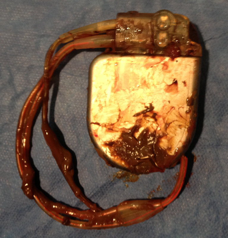
click to enlarge
Questions:
- How do you explain the startling appearance of the removed device?
- With the “muddied” device in your hand, what is your next step in caring for this patient?
- What is the likelihood of infection in this scenario?
- What insights, if any, do clinical guidelines offer for handling a situation like this
Response:
July 24, 2013
1. The appearance is strongly indicative of some type of infection, although atypical for a bacterial process. A reader has suggested a parasitic infection, which is an interesting possibility. Benign processes are almost uniformly serosanguinous; this appearance is quite different. The lack of systemic signs and symptoms does not dissuade me from strongly considering infection; many cardiac device infections do not result in systemic illness.
2. With the device in hand, I would ask that Gram stain and cultures be performed from the device. Blood cultures should be done as well. The entire device should be sent to pathology; a sample of the thickened capsule should also be sent for pathology review. I would consider explanting the just-implanted device, as that would be much easier to do now than later. His arrhythmic risk could be covered with use of a LifeVest (see Chung MK et al., JACC 2010; 56:194). The original indication for the ICD and whether there have been events in the interim are not clear but would affect the decision about whether to use a LifeVest.
3. I think the risk for a localized infection is significant but needs confirmation.
4. The 2010 AHA Update on Cardiovascular Implantable Device Infections is an excellent resource for all physicians who care for such patients. The take-home message is to have a high level of suspicion and to treat aggressively.
Follow-Up:
Westby G. Fisher, MD, and Edward Janszen Schloss, MD
July 31, 2013
Gram stain of the debris showed no white cells or organisms. The capsule was removed in its entirety. Cultures of the pocket capsule tissue, blood cultures, and cultures of the fluid all came back showing no growth. The patient had been on warfarin (and later rivaroxaban) for his atrial fibrillation.
Of course, at the time of the procedure, the culture results were not known. So the new ICD pocket was re-draped and prepped, and the wound was opened to find the tunneled 0074 ICD lead’s tie-down sleeve. This was released, the lead was cut and capped, and the distal portion was withdrawn back to the abdominal pocket to prevent a “wick” extending from the abdominal wound to the new ICD system. The new ICD wound was re-closed.
The abdominal would was irrigated with antibiotic saline solution and closed primarily, and a Jackson-Pratt drain was placed to remove dead space and prevent seroma formation. The antibiotic was expanded to vancomycin only (from cefazolin), continued for 3 days, and then discontinued when the Jackson-Pratt drain was removed.
The patient was seen by an ID specialist, who agreed that stopping antibiotics and observing was the best scenario. The patient was seen in follow-up 10 days later and has done well.
Note that this case was first published on my blog (Dr. Wes), and I would like to credit Dr. Edward Schloss for coming up with the correct answer before I revealed it. Here is an excerpt from Dr. Schloss’ astute response on my blog:
“I’ve come across this exact situation 3 or 4 times before. We have seen this in second or later pulse-generator changes and more commonly in abdominal pockets.
“When an ICD is replaced into a chronic ICD pocket, the remaining open space in the pocket can serve as a cavity that can collect debris. If the pocket capsule is not dissected away partially or completely, the closed pocket will act as a protected closed space. Any remaining blood from the prior surgery will have no place to go. As the generator rubs in the interior of the capsule, the capsule material will slough off and collect in the pocket. Because there is no communication with the subcutaneous space, the debris will simply fill the additional space. The debris is not infectious.
“This is not seen at the time of first pulse-generator changes, because the pocket forms around the new device as the remaining surgical blood drains away. I suspect it happens less often in pectoral pockets, because typically we have to dissect the pocket at least a bit to fit the new device in place. I think any form of extrapocket communication with the subcutaneous tissue will prevent the process. In addition, these devices generally don’t get banged around as much as the abdominal devices.
“Nowadays, I never close a chronic device pocket without at least some form or capsulotomy. So far, I have not seen this happen again.”