
July 17th, 2013
A Surprising ICD Revision
Westby G Fisher, MD, James Fang, MD and Edward J. Schloss, MD
Editor’s Note: This post includes a photo that some readers may find disturbing. The patient has provided consent for the details of his case to be published.
In 1995, a 52-year-old man with chronic atrial fibrillation underwent implantation of an abdominal ICD. As was customary then, the large ICD pulse generator was implanted in the left upper quadrant of the abdomen and connected to an old Guidant Endotak Model 0074 lead that was implanted via the left subclavian vein and then tunneled down to the abdominal pocket. The device served the patient well for years, until its battery depleted in 2003. The old device was replaced by a new, smaller ICD with an appropriate header, and because the defibrillator lead worked well, the smaller ICD pulse generator was left in the abdominal pocket.
Until 2012, the patient underwent routine defibrillator checks in the device clinic without incident. That year, although the lead impedance and capture thresholds remained normal, intermittent periods of noise with nonphysiologic short RR intervals (suggestive of possible impending lead fracture) began to appear on the device checks. The patient was not pacemaker-dependent, so the electrophysiologist decided to wait until the battery would need to be replaced (7 months later) before revising the patient’s ICD system. The EP reasoned that at that time a new defibrillator lead could be implanted and connected to a more conventional VVIR ICD pulse generator implanted in the upper chest area. The old pulse generator could then be removed from the abdomen, and the old lead would be capped and left in place.
The day for surgery arrived. The patient, now 70 years old, felt fine: no fever, chills, or other unusual preoperative symptoms. A venogram performed immediately before the procedure revealed patent left-axillary and subclavian veins. So the decision was made to first implant the new ICD on the same side where the patient’s original defibrillator was implanted, then to remove the old ICD pulse generator from the abdomen. Preoperative antibiotics were administered, and the new single-chamber ICD was implanted via the left-axillary approach without a hitch. A dressing was applied to the wound, and preparations were made to explant the abdominal pulse generator.
The lower abdominal area was similarly prepped and draped. Local anesthetic was infiltrated over the prior abdominal scan, and an incision was made at this location. By means of electrocautery dissection, the incision was carried to the pulse generator capsule, which appeared to be quite thick but not inflamed. The fibrous capsule surrounding the pulse generator was then opened. What was found was startling.
Inside the pulse generator pocket was the device and lead system, surrounded by thick, odorless fluid that looked, for lack of a better phrase, like wet, brown mud. After the suture holding the device’s header to the pocket wall was cut, the device was extracted from the pocket. A portion of the lead was also cut and removed. Here is a picture of the removed device:
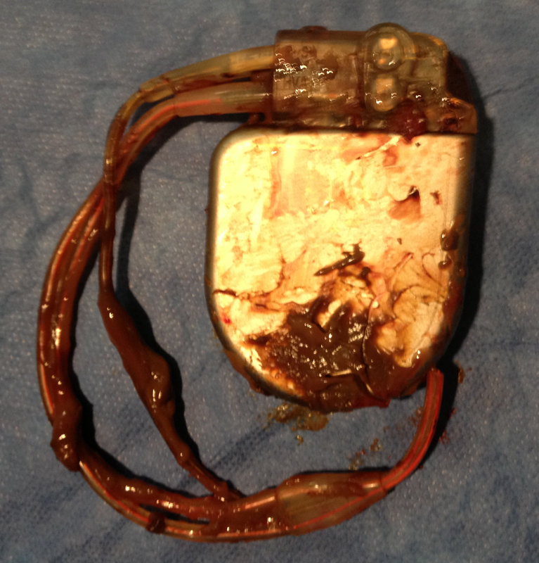
click to enlarge
Questions:
- How do you explain the startling appearance of the removed device?
- With the “muddied” device in your hand, what is your next step in caring for this patient?
- What is the likelihood of infection in this scenario?
- What insights, if any, do clinical guidelines offer for handling a situation like this
Response:
July 24, 2013
1. The appearance is strongly indicative of some type of infection, although atypical for a bacterial process. A reader has suggested a parasitic infection, which is an interesting possibility. Benign processes are almost uniformly serosanguinous; this appearance is quite different. The lack of systemic signs and symptoms does not dissuade me from strongly considering infection; many cardiac device infections do not result in systemic illness.
2. With the device in hand, I would ask that Gram stain and cultures be performed from the device. Blood cultures should be done as well. The entire device should be sent to pathology; a sample of the thickened capsule should also be sent for pathology review. I would consider explanting the just-implanted device, as that would be much easier to do now than later. His arrhythmic risk could be covered with use of a LifeVest (see Chung MK et al., JACC 2010; 56:194). The original indication for the ICD and whether there have been events in the interim are not clear but would affect the decision about whether to use a LifeVest.
3. I think the risk for a localized infection is significant but needs confirmation.
4. The 2010 AHA Update on Cardiovascular Implantable Device Infections is an excellent resource for all physicians who care for such patients. The take-home message is to have a high level of suspicion and to treat aggressively.
Follow-Up:
Westby G. Fisher, MD, and Edward Janszen Schloss, MD
July 31, 2013
Gram stain of the debris showed no white cells or organisms. The capsule was removed in its entirety. Cultures of the pocket capsule tissue, blood cultures, and cultures of the fluid all came back showing no growth. The patient had been on warfarin (and later rivaroxaban) for his atrial fibrillation.
Of course, at the time of the procedure, the culture results were not known. So the new ICD pocket was re-draped and prepped, and the wound was opened to find the tunneled 0074 ICD lead’s tie-down sleeve. This was released, the lead was cut and capped, and the distal portion was withdrawn back to the abdominal pocket to prevent a “wick” extending from the abdominal wound to the new ICD system. The new ICD wound was re-closed.
The abdominal would was irrigated with antibiotic saline solution and closed primarily, and a Jackson-Pratt drain was placed to remove dead space and prevent seroma formation. The antibiotic was expanded to vancomycin only (from cefazolin), continued for 3 days, and then discontinued when the Jackson-Pratt drain was removed.
The patient was seen by an ID specialist, who agreed that stopping antibiotics and observing was the best scenario. The patient was seen in follow-up 10 days later and has done well.
Note that this case was first published on my blog (Dr. Wes), and I would like to credit Dr. Edward Schloss for coming up with the correct answer before I revealed it. Here is an excerpt from Dr. Schloss’ astute response on my blog:
“I’ve come across this exact situation 3 or 4 times before. We have seen this in second or later pulse-generator changes and more commonly in abdominal pockets.
“When an ICD is replaced into a chronic ICD pocket, the remaining open space in the pocket can serve as a cavity that can collect debris. If the pocket capsule is not dissected away partially or completely, the closed pocket will act as a protected closed space. Any remaining blood from the prior surgery will have no place to go. As the generator rubs in the interior of the capsule, the capsule material will slough off and collect in the pocket. Because there is no communication with the subcutaneous space, the debris will simply fill the additional space. The debris is not infectious.
“This is not seen at the time of first pulse-generator changes, because the pocket forms around the new device as the remaining surgical blood drains away. I suspect it happens less often in pectoral pockets, because typically we have to dissect the pocket at least a bit to fit the new device in place. I think any form of extrapocket communication with the subcutaneous tissue will prevent the process. In addition, these devices generally don’t get banged around as much as the abdominal devices.
“Nowadays, I never close a chronic device pocket without at least some form or capsulotomy. So far, I have not seen this happen again.”
I have seen something very similar. The patient was almost asymptomatic and had a quite thick capsule, with a brown thick odorless high viscosity fluid. The difference, it was located in the liver and it was pus from a chronic ameboid liver abscess. Maybe the device was compromise with Entamoeba histolytica.
I want to add the diagnosis of extra intestinal disease is accomplish with serological test. This presentation occur after months or year after exposure, typically you don’t find ova or parasites in the stools. Use a Lugol’s iodine tension to increase detection yield. The fluid usually only have neutrophils.
Possibly an unusual infection? Resolving hematoma from trauma – (could explain the lead issues)?
I would send to pathology. Culture the fluid / send the fluid to pathology for cytology/cell counts etc.
Very interesting case; I think there are several factors to consider.
1) He already has a new device in place; The current issues with pocket were localized not systemic. It could be an indolent infection but more likely a seroma due to placement of a much smaller pulse generator in place of a large generator at the time of last Gen change. If it is a seroma , I think pocket debridment with removal of scar tissue should help resolve any future issues at that site.
2) Worse case scenario, this is an infection; How did it remain localized for years. if it so indolent it may never affect the new device, as it is in different place physically.
3) If one would have seen this finding prior to reimplant, knowing original implant indications would be helpful. He received ICD in early ICD era….so must have had a secondary prevention indication (though I may be wrong in assuming that). Has he ever had any appropriate therapy. We do know that he is not pacing dependent. If has never made good use of his ICD in 18 years, may he doesn’t need another one and we could have done watchful waiting. The question of old lead remains…it is quite high risk for extraction and without a real solid indication for extraction, I am not sure risks will be worth taking it.
I would have performed a full capsulectomy , sent cultures and leave the current device in place unless there is clear evidence of infection.
I will be interested to see the final outcome.
I performed a box change earlier this year on a subpectoral device. The patient was fine, with no issues going into surgery. Dissecting down I was surprised to see the capsule protruding through the pectoral muscle and on opening the capsule there was a large quantity of brown, thick serous fluid. I sent this for cultures and grew nothing.
I was really not sure what to do, but washed the wound out and reimplanted a new ICD. 2 weeks later he represented with wound dehiscence and we extracted the system.
I am not sure if there was latent infection there, or whether I introduced infection. But latent, asymptomatic infections do seem to be described in the literature.
I suspect this was a chronic indolent infection confined to the pocket/capsule. The concern will be whether now a systemic infection will occur since the pocket/capsule has been manipulated. Nevertheless I would not explant the pectoral device. I’d place the patient on antibiotics and follow closely. Presuming the reason for implant originally was appropriate. I’d also get an abdominal x-ray to be sure there is no sub-diphragmatic air, and consider a search for a sub-diaphragmatic abscess. Although lack of systemic findings makes this unlikely.
My experience with the Lifevest is that the noncompliance rate is quite high, hence I’d stay with the pectoral implant. If infection occurs later, of course entire removal will then be necessary.
This is a fascinating case, very rare. The hypotheses of that a remaining open space in the pocket, can serve as a cavity that can collect debris and produce a thick, odorless fluid that looked, for lack of a better phrase, like wet, brown mud is an interesting possibility.
Dr. Westby G. Fisher, MD, and Edward Janszen Schloss, MD could you please elaborate more? How did you test and confirm this plausible conjecture or hypotheses? At this moment the diagnosis is very important but taking in consideration how rare this is it. I am more interested in the process of How do you arrive at this final conclusion? Do you have objective confirmation? Is this a exclusion diagnosis?
Dr. Guadiana,
Than you for your interest in this case. My reply to Dr. Fisher’s case originally went up on his blog. My comments were based on personal experience and not rigorously validated. My conclusion of the mechanism is based on logic, and I invite other explanations of our findings. That said, we have never seen any long term issues with these cases. I’ve had two or three myself that did not get antibiotics and one other case locally did well, although I was not involved directly.
If I were a better academician, I would have collected and reported the data systematically, and I now regret that I did not. My hope is that we will hear of other’s experiences through social channels such as Cardioexchange.
Jay
Edward J. Schloss MD FACC FHRS
Medical Director, Cardiac Electrophysiology
The Christ Hospital
Cincinnati, OH
USA
schlossj@ohioheart.org
@EJSMD
Dr. Gaudiana –
Because this finding is relatively rare and has been seen be several of us, we can only surmise the mechanism. As you suggest, it is a diagnosis of exclusion. We performed gram stains, cultures for bacteria and fungus, and sent pathology and cultures of the fibrous ICD capsule – all of which failed to disclose a pathologic organism. The lack of white cells or organisms within the “mud”-like material, paired with lack of fever and signs of infection, added to the clinical picture of a relatively benign process.
After removing the abdominal ICD pocket and evaluating the patient one month later, he has done well and his cultures never grew any organism.
To me, cases like this make a strong case for social media in medicine. We have seen large peer-reviewed journals often excluding case reports for publication due to the cost to publish cases like these and the desire to improve their impact factors which are strengthened by randomized clinical trial publications. As such, I appreciate the editors of this blog sharing this case with their readership.
Thank you very much Dr. Schloss and Dr. Fisher I appreciate your response and I fully agree with both of you. Without a doubt social media in medicine have revolutionized the exchange of knowledge specially with participation of excellent physicians and academics like the former colleagues and the impact in the practice of medicine is enormous.
I believe, in cases like this, the abstraction process and how we test our hypotheses makes us more respectful of how difficult is to arrive at a diagnosis and generate knowledge and indisputably help us to understand
An inquisitive approach in cases like this is the best.
Thank you for sharing this interesting case and your experiences.