
April 10th, 2017
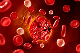
Pulmonary Embolus: Evaluating the Five Ps of PE
Alexandra Godfrey, BSc PT, MS PA-C

Alexandra Godfrey, PA-C, practices emergency medicine in North Carolina.
Years ago, my physician father said to me; “If something does not make sense, if you struggle to determine the pathology, consider pulmonary embolus (PE).”
More recently, an ER physician colleague offered me the following advice: “If you think about PE, test for it.”
Of course, an astute clinician places both pieces of wisdom into context. I am not going to look for PE in patients presenting with ambiguous abdominal pain nor am I going to order d-dimers on all patients with a hint of PE. I also accept that tucked in the fables and parables of medicine are certain truths that deserve my attention. The first piece of advice speaks to the cryptic and non-specific presentation of pulmonary embolus and the second refers to the importance of clinical suspicion or gestalt.
The mortality, morbidity, and prevalence of PE combined with its enigmatic presentation can cause trepidation and uncertainty in providers, sometimes triggering haphazard or inappropriate testing. Such testing may result in poor outcomes for patients (and providers). Moreover, the confusion is compounded by conflicting opinion and divergent practice: some providers never order a d-dimer, some order one on every low-risk patient, some start with CTA, etc. If I ask three clinicians about their work-ups, I will likely get three quite different answers. Furthermore, anecdotes are powerful; we all have stories about the most unlikely presentation of PE. Consequently, PE evaluation can be as murky and off-putting as my grandmother’s pea soup (sorry, grandma). Nonetheless, even though PE is a pernicious, petulant and peculiar pathology, we can find clarity if we carefully consider the five Ps of PE evaluation. What is more – you can count them on the phalanges of one hand.
P1. Patient Presentation
Step one: look at your patient. Prior to any testing, perform a thorough history and physical. Patients with PE present in a variety of ways and symptoms can be subtle. Symptoms of PE include: dyspnea, pleuritic chest pain, cough, syncope, hemoptysis, tachycardia, fever, leg swelling. Dyspnea and chest pain are the most common symptoms of PE; however, neither is sufficiently sensitive or specific to rule pulmonary emboli in or out.
P2. Pre-test Probability
Step two: determine pre-test probability. You can use either your gestalt or validated clinical prediction rules to further stratify risk in your patient. My preference is to use a combination of the two, as my years in medicine have taught me two facts: firstly, my judgement is fallible, and secondly, some patients do not fit the rules. Knowing in medicine is never a simple algorithm or a score; it is a shared multidimensional process that transcends these.
P3. Prediction Rules
Step three: prediction rules. Well validated clinical prediction tools include the revised Geneva (rGeneva) and the two-step Wells’ scores.1 The rGeneva score is entirely objective. Patients are stratified in low, intermediate and high risk. The two-step Wells’ score divides patients into PE unlikely (≤4) and PE likely (>4). The criticism of the Wells’ score is that it incorporates clinical gestalt by giving 3 points for suspicion for PE. Both scores guide subsequent testing. A patient who is <50 years old and is considered low risk by gestalt or prediction tool can be further ruled out by using the Pulmonary Embolism Rule Out Criteria (PERC).
Undoubtedly, given the variability in use and inconsistent knowledge of these rules, serious consideration should be given to incorporating clinical decision tools (as computer support aids) in electronic charting, especially for providers who are inexperienced or risk-averse, or who infrequently stratify PE risk. These aids do not usurp gestalt but rather enhance it.
 P4. Pesky D-Dimer
Step four: the d-dimer. Patients who don’t meet PERC should be further stratified by the plasma d-dimer, a fibrin degradation product that when present indicates the initiation of procoagulant and fibrinolytic pathways. D-dimers may rule out PE in low-risk and even moderate-risk patients, depending on the assay used. Remember, they are sensitive but not specific. Moreover, do not order d-dimers on high-risk patients as the increased incidence of disease in these patients creates spectrum bias (if you change the patient case mix, you change the performance of a test).
Different assays are available and have different cutoffs and units. A common and sensitive d-dimer assay is the ELISA. Another sensitive assay is the immunoturbidimetric. Both are quantitative. One of my practice sites uses the former and the other uses the latter. If you plan to age adjust the d-dimer you must know the following i) the type of assay, ii) the reporting unit, and iii) the cutoff. If you don’t know these things, you could make critical errors.
The greatest use of the d-dimer stems from its high negative predictive value. Thus the d-dimer is for patients who are “PE unlikely” by Wells (≤4) or gestalt but are not ruled out by PERC. The ELISA d-dimer assay can also be used in conjunction with clinical prediction tools to rule out patients with intermediate risk.
Age adjusting the d-dimer to further stratify risk and reduce unnecessary imaging while maintaining sensitivity is a hot topic. The American College of Physicians recommends using age-adjusted d-dimer thresholds (age x 10 ng/ml) rather than a generic cutoff to determine d-dimer elevation in patients older than 50 years. Most faculty I asked stated that they would support use of age-adjusted d-dimers in PE algorithms, despite some doubts regarding population cohorts, gold standards, and variant assays. If you want to delve further into the potential problems and pitfalls of the d-dimer, Rory Spiegal MD, provides an interesting discussion of these in his blog
I consider some practices to be poor: for example, ordering d-dimers in triage. Typically, triage is not conducive to a thorough exam nor does it give time for accurate risk assessment; such profligate ordering might incite the wrath of your colleagues. I made this error myself as a new grad PA in triage. Lesson learned. If you see this, kindly put the kibosh on it. Furthermore, the d-dimer isn’t a lab you order and then fail to follow up. Just don’t do it. Indiscriminate use of the d-dimer jeopardizes the safety of your patient whereas judicious use protects your patient.
P5. Pictures
Step 5: Imaging studies. Patients with a positive d-dimer or high pre-test probability require imaging studies. The gold standard is the CTA PE. This test is not innocuous. It may detect subclinical PEs, result in contrast-induced nephropathy, cause allergies, and create a financial burden for the patient. However, in the high-risk patient, the CTA is the go-to test. VQ scanning is useful for patients with contraindications to CT but not for patients with CHF, COPD or pneumonia. Another valid strategy is to order venous duplex studies of the lower extremities prior to CT as this may eliminate the need for CT and obviate its inherent risks.
In conclusion, pernicious, petulant, and peculiar is as pernicious, petulant and peculiar does; yet by paying attention to the Ps of PE evaluation, you can improve outcomes, reduce cost, sharpen your skills as a diagnostician, and enjoy your own brand of P soup.

Reference
-
Douma RA et al. Prometheus Study Group. Performance of 4 clinical decision rules in the diagnostic management of acute pulmonary embolism: a prospective cohort study. Ann Intern Med. 2011; 154:7009.

15 Responses to “Pulmonary Embolus: Evaluating the Five Ps of PE”

NP/PA Bloggers
Elizabeth Donahue, RN, MSN, NP‑C
Alexandra Godfrey, BSc PT, MS PA‑C
Emily F. Moore, RN, MSN, CPNP‑PC, CCRN
Advanced practice clinicians treating patients in a variety of settings and specialties
Learn more about In Practice: Reflections from NPs and PAs.
Are you following the international accepted Abbreviation list and I can look up these abb’s (abbreviations)?
P.S. Read hospital discharge note recently describing patient’s “OP” (the treatment worthy disease ‘osteoporosis’ the Medical Records Department assured me – only problem – patient actually had the non-treatment worthy state of osteopenia (“OP”) when the dictating physician was eventually traced.
Great pea soup – no dead pig in it I hope.
Good general medical (thought) algorithm – could be expanded to alcoholism, Tuberculosis, myocardial infarction, etc.
Hi Max –
I laughed at your comment about abbreviations. So true; there’s so many of them and they are often singular to the individual or specialty. Usually best to just write it out if in doubt.
Yes, you are correct – you could probably apply this to many presentations in medicine.
No bacon on this pea soup but it would make it more tasty perhaps.
I like your suggested approach. The only question I had relates to the use of the term “subclinical” PE. I know that sometimes we find incidental PE if we give contrast for another reason but if you are referring to patients who underwent a chest CT wiht PE protocol then there must have been a clinical reason to give contrast-in that case the PEs could not have been “subclinical”.
Andre -you are correct. Subclinical or incidental PE is usually a finding on imaging that is done for other reasons, such as on a CT chest to evaluate a pulmonary nodule or some such thing. The suspicion here not being for PE and the PE being an incidental finding. And of course a standard CT chest may produce a false positive due to the different timing of the contrast.
More accurate terms might be “clinically irrelevant” or “clinically insignificant clot burden”. It refers to the tendency to find pathology where perhaps there isn’t one or at least begs the question – are the risks of anticoagulation in these patients greater than the risks from tiny sub-segmental PEs? Are these always pathological? Of course, this is a dicey area as what might be clinically significant in one patient may not be in another, and I am not sure we have parameters that determine this well.
In PIOPED II, the largest trial examining the diagnostic characteristics of CTPA, Stein et al found that in patients with low-risk of pulmonary embolism by clinical assessment, the CTPA diagnosed far more PEs than the composite reference standard (Normal DSA or V/Q scan, a low probability V/Q with Wells <2 or a negative LE US). Moreover, 42% of the PEs diagnosed by CTPA were false positive findings. This suggests we should be cautious about scanning these patients and rely more on prediction rules, probability and gestalt. If we go straight to CTA in low risk patients, we could be anti-coagulating patients with false positives. Additionally, if a patient is low risk could we consider their PE on CT to be subclinical, meaning does just the thought of PE (clinical suspicion worth 3 points on Wells) make the pathology clinically evident?
Thank you for picking up on this.
As a psychiatrist, I appreciate your logical reasoning and your wonderful writing, even if I am not trying to detect PEs in the ED. I think your decision making approach can be applied to many different situations.
I will looking for more of your articles in the future!
Thank you,
Elias
PS. I wish I could still get some of my mom’s split pea soup. It was wonderful.
Elias H Sarkis, MD
Gainesville, FL
Thank you Elias. I am glad you enjoyed it. And I agree, this type of thinking could be applied to many different situations. It sounds like your mom was better at pea soup than my grandma, but to be fair, she did make a fine upside down almond pudding.
Thank you, Alex! Great article!
I have not been using PREC and will be looking forward to applying this approach in working up for underlying PE.
Well written article. If you would have made a flow chart and included that would be a good summary.
Thank you for the suggestion Abdul. You are correct; a flow chart would have been a good addition to this article. Next time, I write something similar I will consider it. I hope this approach serves you well.
Discourage ordering d-dimer in post-operative patients. It will give you a (possibly false) positive result. Because these patients are often less mobile, their risk for DVT and PE is higher. Forget the d-dimer and go straight to CTA
I agree with this. They are not low risk by virtue of recent surgery and immobilization. If your clinical suspicion is high, and your pre-test probability is high, then CTA is the answer.
The biggest risk of the CTPA isn’t cost (minimal), time (not insignificant), contrast nephropathy (does it really exist), placing an unwell or in a high ridk environment (the CT scan), radiation (minimal), but false positive scans. With improved CT technology increasing sensitivity, there are inevitably more false positives. So what does this mean ….. injections, daily tablet for at least 3 months, increased risk of bleeding etc. but also the hidden burden- falsely having a history of PE- which influences every operation from then on, every chest pain or dyspnoea presentation (tick prior PE/DVT) etc. the non-financial costs are indeed non insignificant
Paddy – you raise a very valid point. Having a false positive brings unnecessary treatment and an inaccurate assessment of future risk and with that more unnecessary treatment. Thank you for your comment. I think you are correct in your concern.
In my exprience had very diverse presentations of suspicious or definite PTE from typical symptomatic pt with neg CTA &ordinary respiratory symptoms such as cough that we hadnt any doubt to PTE but pt had cardiopulmonary arrest that didnt respond to CPR so we must always consider this Dx but also dont treat pt with anticoagulant who doesnt need it.
A excellent summary of PE workup. Next question. How do we decide to treat or not treat sub segmental PE. And what guidelines should we be using to manage submissive PE.
The American College of Physician Review in 2015 is excellent.. This 5P’s is consistent. For those of you who have not read the ACP publication.
The review was published at http://www.annals.org on Sept 29, 2015 for those you are interested. I highly recommend it.